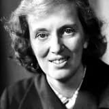
With J.D. Bernal, Hodgkin showed X-ray diffraction patterns required hydrated protein crystals, a founding insight of protein crystallography.
In 1934, working alongside her mentor J.D. Bernal at Cambridge, Dorothy Hodgkin co-authored a landmark paper demonstrating that meaningful X-ray diffraction patterns could only be obtained from protein crystals kept in their natural, hydrated state, rather than dried crystals as had been common practice.
This discovery was pivotal: previous attempts to study proteins by X-ray crystallography had produced poor or misleading results because drying crystals disrupted their internal molecular order. Bernal and Hodgkin's insight—that proteins needed to remain in mother liquor during X-ray exposure—instantly improved the quality and reliability of protein diffraction data across the field.
The publication, which reported some of the first clear X-ray diffraction images of the enzyme pepsin, is widely regarded as a founding milestone of modern protein crystallography, a discipline that would eventually reveal the atomic structures of DNA, hemoglobin, and thousands of other biological macromolecules.
For Hodgkin, still in the early stages of her career, this work established her reputation as a serious experimental scientist and gave her the technical grounding she would later apply to penicillin, vitamin B12, and insulin. It also cemented a lifelong collaborative and intellectual relationship with Bernal, one of the most influential crystallographers of the twentieth century.